Description
A 79-year-old Thai man with a history of untreated mid-oesophageal cancer presented with progressive dyspnoea and productive cough for 3 weeks. On examination, he had low-grade fever, tachyponea, regular pulse rate at 110/min, blood pressure 120/78 mm Hg without definite pulsus paradoxus. Pulmonary examination showed mild deviation of trachea to the right, dullness on percussion and decreased breath sound at the left lung. He also had mildly distended neck veins, decreased cardiac sound and decreased apical impulse. ECG revealed sinus tachycardia with low QRS voltage without significant ST-T change. A large amount of generalised high-echoic pericardial fluid and evidences of early cardiac temponade were demonstrated by transthoracic echocardiography.
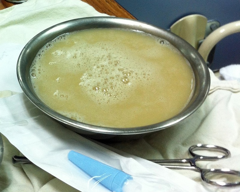
Contrast-enhanced CT of the chest showed known circumferential oesophageal mass at the middle one-third of the oesophagus (black asterisk, figure 1A). A large amount of circumferential pericardial fluid (white asterisks, figure 1A–C) with small air bubbles (bold arrows, figure 1A–C) were detected. In addition, large loculated fluid collections in left hemithorax with air-fluid level and pleural enhancement (open arrows, figure 1A,C) were observed. Subsequent pericardiocentesis along with left intercostal drainage were performed, which revealed similar findings of large volume of frank, yellowish, foul-smelling pus (figure 2), consistent with pyopneumopericardium and empyema thoracis. On the basis of these findings, the patient underwent surgery with the presumptive diagnosis of the perforation of necrotic oesophagus leading to direct spread of infection to adjacent organs. Intraoperative finding confirmed the ruptured oesophagus at mid-thoracic level as a consequence of local tumour invasion. He successfully underwent combined drainage of pus in pericardial space with pericardiectomy, decortication of left lung and resection of the thoracic oesophagus from just below aortic arch to oesophagogastric junction. Microbiological cultures of pus were positive for mixed organisms including Pseudomonas aueruginosa, Streptococcus group D and Streptococcus group G. Postoperative period was uneventful and he is currently in the restoration process of enteral alimentation before being discharged.
Figure 1.

Contrast-enhanced CT in axial (A), coronal (B) and sagittal views (C) show malignant oesophageal mass encasing the nasogastric tube at mid-oesophagus (black asterisk, A). A large amount of circumferential pericardial effusion (white asterisks, A–C) with small air bubbles (bold arrows, A–C) indicated pyopneumopericardium. Large loculated fluid collections in left hemithorax with air-fluid level and pleural enhancement of its thin wall (open arrows, A and C) represented empyema thoracis.
Figure 2.

Frank, yellowish, foul-smelling pus from pericardiocentesis which appeared similar to empyema thoracis from intercostal drainage. Microbiological culture was positive for mixed organisms including Pseudomonas aueruginosa, Streptococcus group D and Streptococcus group G.
Perforation of oesophageal cancer is a serious circumstance that most often results from either diagnostic or therapeutic instrumentation, that is, stent placement.1 Locally advanced cancer may lead to spontaneous perforation of oesophagus which has high mortality rate.2 3 This allows progressive migration of the normal bacterial flora from the oesophagus to adjacent mediastinal organs include of the pericardial sac and pleural cavity leading to uncontrolled infection.4 Pyopneumopericardium and empyema thoracis are very rare, but recognised, complications which dictate prompt surgical therapy.
Learning points.
Perforation of oesophageal cancer is a serious circumstance that most often results from either diagnostic or therapeutic instrumentation.
Advanced oesophageal cancer may lead to spontaneous perforation from local tumour invasion.
Pyopneumopericardium and empyema thoracis are very rare complications as a result of progressive migration of the normal bacterial flora from the oesophagus to adjacent mediastinal organs.
Footnotes
Contributors: NC and PC wrote the manuscript. MT, KL reviewed and revised the manuscript.
Competing interests: None.
Patient consent: Obtained.
Provenance and peer review: Not commissioned; externally peer reviewed.
References
- 1.Huber-Lang M, Henne-Bruns D, Schmitz B. Esophageal perforation: principles of diagnosis and surgical management. Surg Today 2006;2013:332–40 [DOI] [PubMed] [Google Scholar]
- 2.Reeder LB, DeFilippi VJ, Ferguson MK. Current results of therapy for oesophageal perforation. Am J Surg 1995;2013:615–17 [DOI] [PubMed] [Google Scholar]
- 3.Bhatia P, Fortin D, Inculet RI, et al. Current concepts in the management of esophageal perforations: a twenty-seven year Canadian experience. Ann Thorac Surg 2011;2013:209–15 [DOI] [PubMed] [Google Scholar]
- 4.Piatkowski R, Kochanowski J, Karpiński G, et al. Purulent pericarditis in patient with esophageal cancer presenting with cardiac tamponade. J Emerg Med 2011;2013:671–3 [DOI] [PubMed] [Google Scholar]


