
Figure 1.
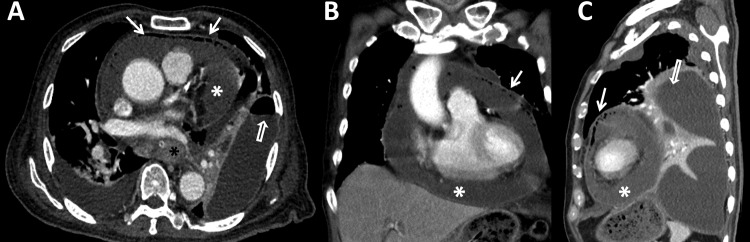
Contrast-enhanced CT in axial (A), coronal (B) and sagittal views (C) show malignant oesophageal mass encasing the nasogastric tube at mid-oesophagus (black asterisk, A). A large amount of circumferential pericardial effusion (white asterisks, A–C) with small air bubbles (bold arrows, A–C) indicated pyopneumopericardium. Large loculated fluid collections in left hemithorax with air-fluid level and pleural enhancement of its thin wall (open arrows, A and C) represented empyema thoracis.