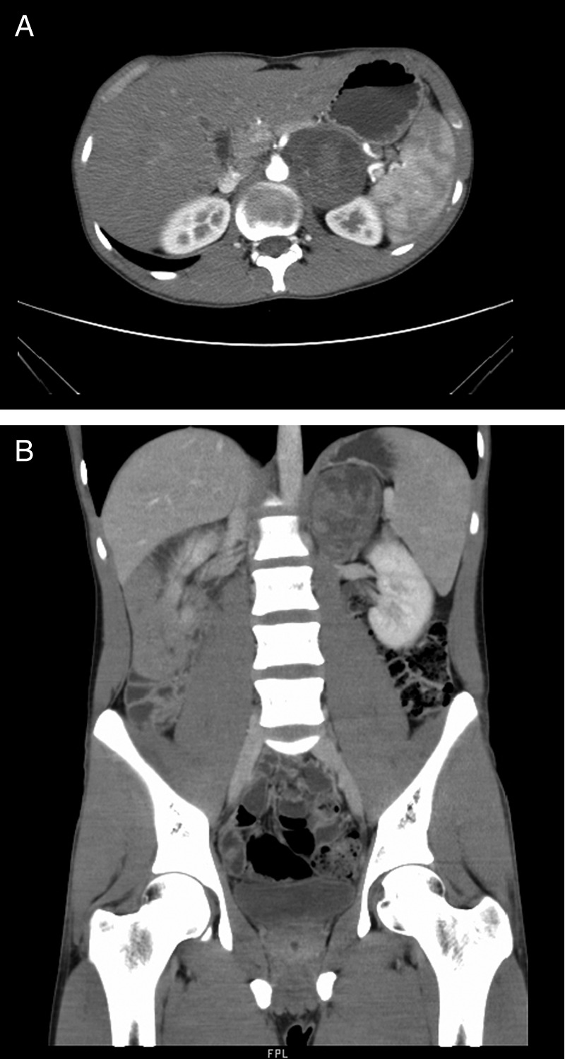
Figure 1.

(A) Contrast enhanced CT (CECT) axial images showing a 6.8 cm×7 cm×5.5 cm well-defined heterogeneous hypodense lesion in the left adrenal gland. (B) CECT coronal images showing a 6.8 cm×7 cm×5.5 cm well-defined heterogeneous hypodense lesion in the left adrenal gland.
