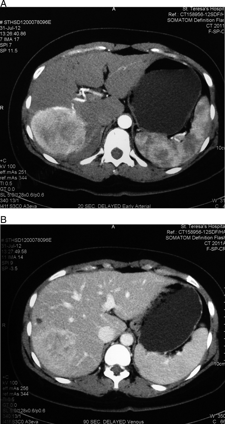
Figure 1.

(A) Contrast CT scan showing a large tumour at the right lobe of the liver with arterial enhancement at postcontrast image. (B) Contrast CT scan showing vague contrast enhancement of tumour with ‘washout’ pattern at 90 s postcontrast image.
