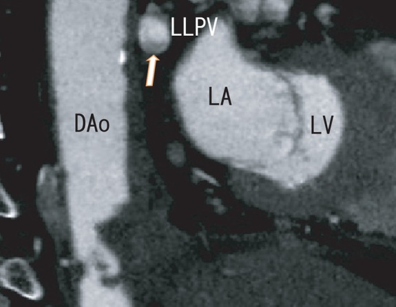
Figure 5.

Sagittal images illustrated the same thrombus in the LLPV as the defect of contrast enhancements (arrow), which became again larger (10×5 mm) and attached to the lower surface of the LLPV. Dao, descending aorta; LA, left atrium; LLPV, left lower pulmonary vein; LV, left ventricle.
