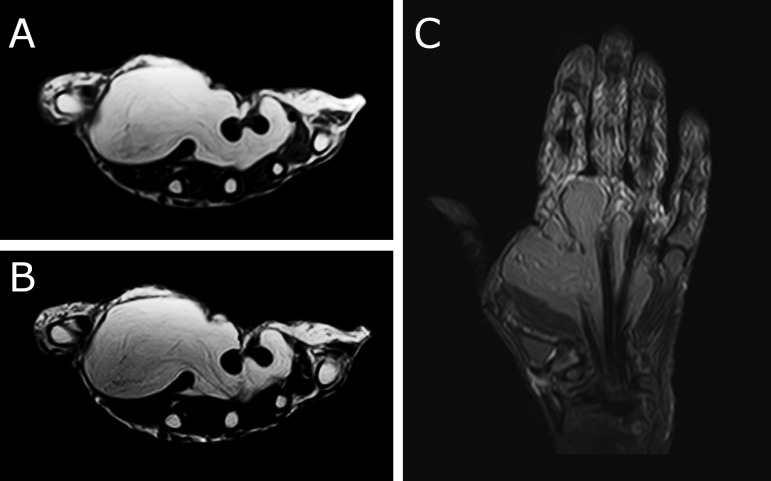
Figure 2.
Axial T1-weighted (A) and T2-weighted (B) images of the left hand showing a large hyperintense soft tissue mass occupying most of the hand, extending from the first to fourth metacarpals (B). On Coronal STIR image (C) it extends from distal carpal row to the metacarpophalangeal joint and shows uniform suppression of signal, indicating its’ fatty nature.

