
Figure 1.
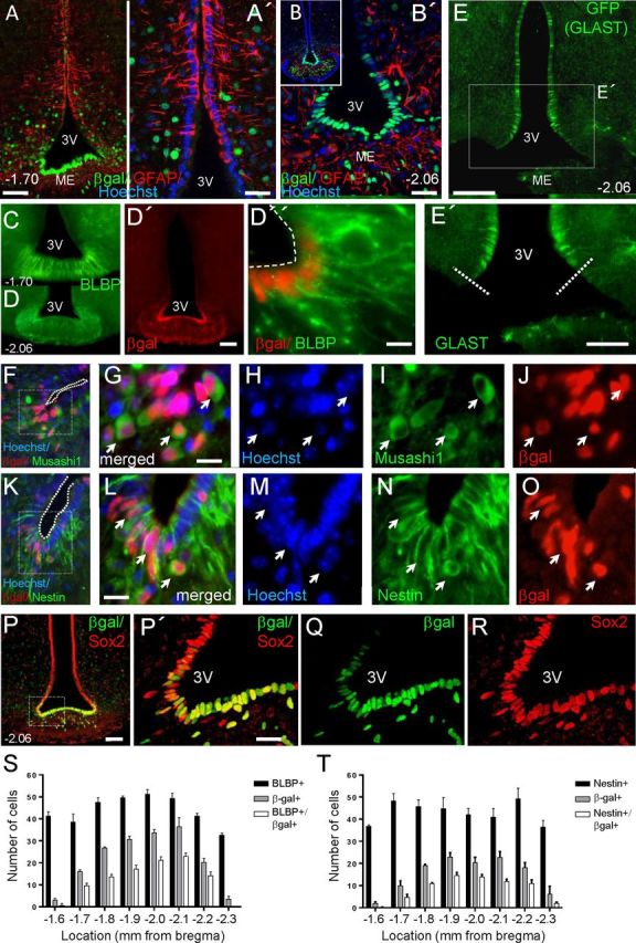
Fgf10+ tanycytes express neural stem/progenitor markers and are most abundant in the central regions of the median eminence. Images show coronal vibratome (A–E', P–R) and cryostat (F–O) sections of P60 Fgf10nlacZ brains, at the level of ME (bregma −1.70 or −2.06, as indicated). Fgf10+ tanycytes, visualized by anti-βgal immunolabeling, coexpress markers of neural stem/progenitor cells: BLBP (C–D”), Musashi1 (F–J), nestin (K–O), and Sox2 (P–R), but not GFAP (A–B'). Note that expression of Sox2 is not limited to ependymal cells, but also present in some parenchymal cells. E, E', Domain of GLAST expression in the 3V ependyma as visualized by anti-GFP immunolabeling of tamoxifen-treated GLASTcreERT2:R26GFP brain. Dashed lines in E' show the ventral limit of GLAST expression. S, T, Fgf10+ tanycytes are more abundant in the central regions of the ME, where only a subpopulation expresses BLBP or nestin. Scale bars: A, B', D', E, P, 50 μm; A', E', P', 25 μm; D”, G, L, 10 μm.