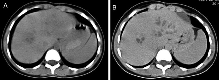
Figure 2.

(A) CT scan of the abdomen showing conglomerate of ill-defined oval cystic lesions along right as well as left hepatic ducts. (B) Contrast-enhanced CT scan of the abdomen in portal venous phase showing slight peripheral enhancement of the walls of the lesions which are now more conspicuous.
