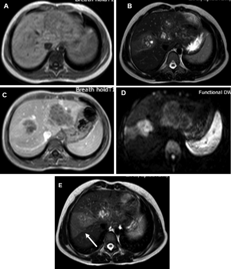
Figure 3.
(A) T1-weighted (T1W) axial MRI image showing the lesions to be hypointense. (B) T2-weighted (T2W) axial image showing the lesions to be hyperintense. (C) T1W contrast-enhanced axial image showing slight peripheral enhancement of the lesions in portal venous phase. (D) Diffusion-weighted image with b value of 1000 showing hyperintensity of the lesions. (E) T2W axial image at slightly higher level showing wedge-shaped area of hyperintensity (arrow). The corresponding areas were hypointense on ADC map (image not available) S/O restricted diffusion.

