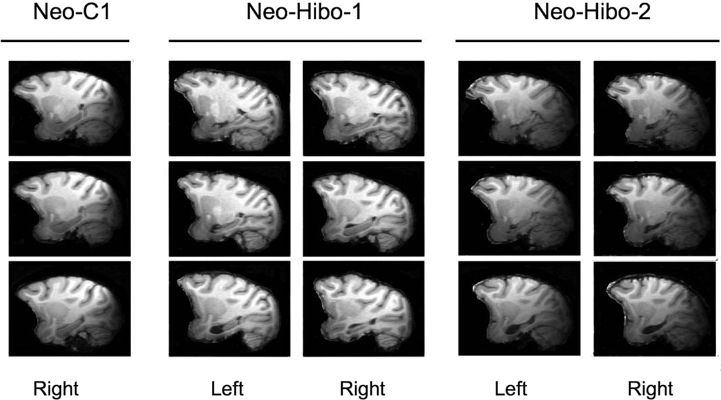
Figure 3.
Magnetic resonance images (T1-weighted), taken in adulthood at 8 to 9 years of age, of one control and two subjects from Group Neo-Hibo at three sagittal levels through the left and right hippocampus. Subject Neo-Hibo-1 has a largely unilateral lesion with more sparing of the right hippocampus (Table 3), whereas subject Neo-Hibo-2 has a bilateral lesion with minimal sparing.

