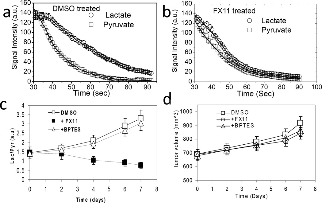
Figure 2.
Tumor [1-13C] lactate and [1-13C] pyruvate peak intensities with time after i.v. injection of hyperpolarized [1-13C] pyruvate in (a) DMSO treated (control) and (b) FX11 treated tumors for 4 days. The initial 30 s of data were not shown because of the time taken to pyruvate deliver and uptake by the tumor. The data were also fitted to two-site exchange model to estimate the rate constants kP and kL. (c) The lactate-to-pyruvate flux ratio (Lac/Pyr) increased with treatment days in DMSO and BPTES treated mice and decreased in FX11 treated mice. (d) A slight increment of tumor volume (measured from T2-weighted MRI) was observed with no significant differences between groups. Error bars are the standard deviation (s.d) from the mean values, n = 8 for each group of mice.

