
Figure 5.
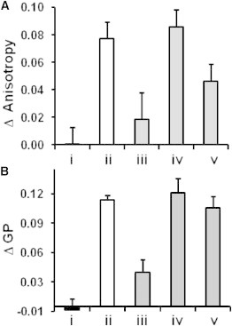
Modulation of bilayer fluidity by β2m amyloid fibrils and different molecules. Changes in (A) fluorescence anisotropy of TMA-DPH and (B) Laurdan emission shift (quantified by GP, Materials and Methods) assayed within PC/PG (1:1) LUVs. The vesicles incubated with (i) β2m monomers, (ii) β2m fibrils, (iii–v) β2m fibrils preincubated with (iii) bromophenol blue, (iv) full-length heparin, and (v) heparin disaccharide before mixing with the vesicles.