Figure 1.
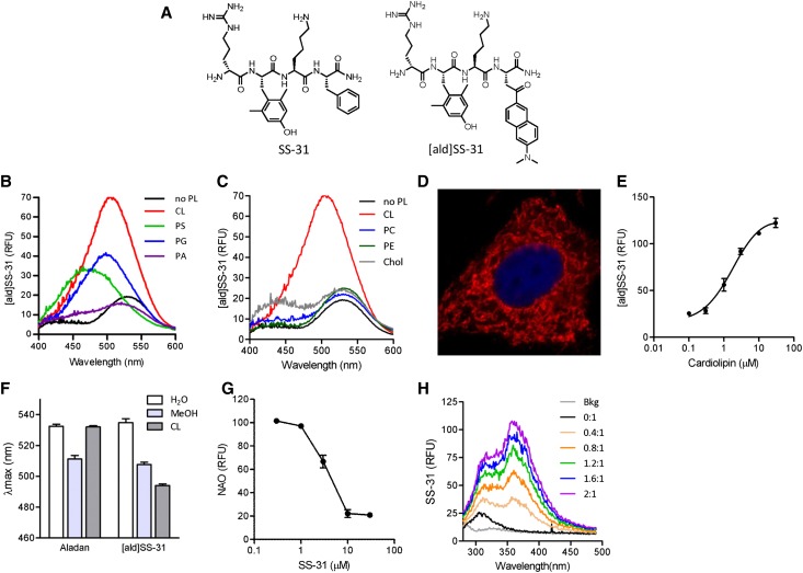
SS-31 interacts selectively with CL. (A) Chemical structures of SS-31 and [ald]SS-31. (B) Representative fluorescence emission spectra of [ald]SS-31 (1 μM, λex=360 nm) in the presence of different anionic phospholipids (3 μM). A shift in emission maximum (λmax) and increase in fluorescence intensity were observed with the addition of CL, PS, and PG but not with PA. (C) Representative fluorescence emission spectra of [ald]SS-31 in the presence of other lipids (3 μM). Chol, cholesterol. There was no shift in λmax with the zwitterionic phospholipids or cholesterol. (D) Intracellular localization of biotinylated SS-31 in feline kidney cells. Biotin was visualized with streptavidin-AlexaFluor594. (E) The interaction of CL with 1 μM [ald]SS-31 is saturable, with KD=1.87±0.64 μM. (F) Representative fluorescence emission spectra of [ald]SS-31 (1 μM) showing a concentration-dependent increase in fluorescence intensity with the addition of CL. (G) SS-31 displaces nonyl acridine orange (NAO) interaction with CL in a competitive manner. (H) Representative fluorescence emission spectra of 10 μM SS-31 (λex=254 nm) with the addition of CL at different CL:SS-31 ratios. RFU, relative fluorescence units.

