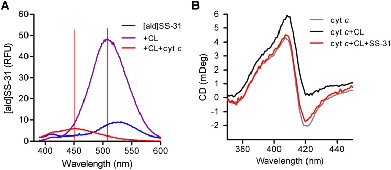
Figure 3.
SS-31 prevents CL from exposing the heme Fe in cyt c to induce peroxidase activity. (A) Change in emission spectrum of [ald]SS-31 on addition of CL and cyt c. Addition of 6 μM cyt c to [ald]SS-02 (1 μM) and CL (3 μM) causes dramatic quenching and additional shift of λmax from 510 to 450 nm, suggesting that the CL–peptide complex resides in a hydrophobic domain in close proximity to the heme. (B) SS-31 prevents the effect of CL on the negative Cotton peak in Soret spectrum of cyt c. CD was carried out with 10 μM cyt c alone (gray) or in the presence of 20 μM CL (black) or CL plus 10 μM SS-31 (red).

