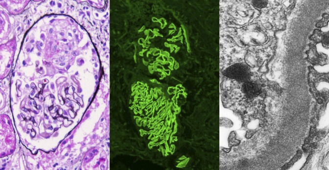
Figure 1.

Crescentic GN with linear GBM staining on immunofluorescence. There is a small cellular crescent with fibrinoid material, with no proliferation or sclerosis of the glomerular tuft (left panel, Jones silver stain; original magnification ×400). By immunofluorescence, there is linear staining along the GBM with antibody to IgG. The top glomerulus also shows a small cellular crescent (middle panel, anti-IgG immunofluorescence; original magnification ×200). By electron microscopy, a high-power view of the capillary wall shows intact foot processes (right), and no deposits were present in a subepithelial or subendothelial location. Reticular aggregates were present in the endothelial cell cytoplasm, consistent with high interferon levels in this HIV-positive patient (transmission electron microscopy; original magnification ×8000).
