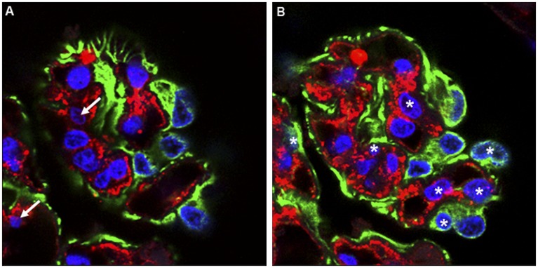
Figure 2.
The physical disector is based on the new appearance of a particle between two physical tissue sections with known separation–implying that the leading point of the particle first occurred within the volume contained between the sections. As long as the specific particle can be identified as present or absent in the two sections, the event of its appearance between the sections is independent of its size or shape and the particle can be unambiguously and uniquely attributed to the volume element in which it first appeared. The disector so described involves two physical tissue sections (called the look-up, here A, and reference section, here B). Notice that in the reference section (B) there are seven appearances of podocyte nuclei (blue) not present in the look-up section (A). When counting cell nuclei with physical disectors, section pairs (A, B) are used to count appearing and disappearing nuclei. In this example, 1-µm confocal optical images separated by 4 µm are used. Paraffin sections from a human glomerulus were stained for Wilms tumor 1 (WT-1) antigen to localize podocytes (green cytoplasm), von Willebrand factor (vWF) to localize endothelial cells (red cytoplasm), and DAPI for all nuclei (blue). Nuclei present in the look-up section (A) that were not present in the reference section (B) were counted (white arrows, two nuclei). Then, those nuclei present in the reference section (B) but not in the look-up section (A) were counted (asterisks, seven nuclei).

