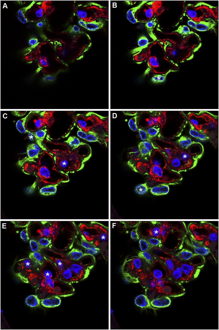
Figure 3.
When cell nuclei are counted with optical disectors, nuclei are counted at a unique moment (e.g., when they first come into focus). In this figure, 14-µm paraffin sections were immunofluorescently stained for Wilms tumor 1 (WT-1) to identify podocytes (green cytoplasm), von Willebrand factor (vWF) to identify endothelial cells (red cytoplasm), and DAPI for all nuclei (blue). Each paraffin section was then optically sectioned using confocal microscopy at 1-µm intervals; six optical sections (A–F) are shown. Nuclei in focus in A were not counted because they did not come into focus. Nuclei that came into focus in sections B–F are indicated by asterisks. The presence of WT-1 and vWF identified which of the counted cells were podocytes (three cells) and which were endothelial cells (seven cells).

