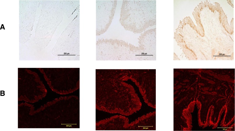
Figure 4.
TNXB is expressed in the UVJ of normal and refluxing ureters. Human UVL sections obtained from normal controls and patients undergoing ureteral re-implantation for radiographically demonstrated PVUR. (A) Immunohistochemistry. Left to right: normal (nonrefluxing ureter) negative control, not stained with rabbit anti-TNXB antibody. There is absence of staining in all layers of the urothelial lining. Second panel: Normal control (nonrefluxing) ureter sample, stained with rabbit anti-TNX antibody (1:50). Note positive (brown) and equal (same intensity) staining throughout all of the urothelium. The third panel represents a refluxing vesicoureteral section of ureter, stained with rabbit anti-TNXB antibody (1:50). Note positive staining of all urothelial layers, but with a stronger intensity staining of the basal layer, compared with the normal control sample in the second panel. (B) Immunofluorescence. Left to right: normal (nonrefluxing ureter) negative control, not stained with rabbit anti-TNXB antibody. There is absence of staining in all layers of the urothelial lining, with nonspecific occasional superficial tissue auto fluorescence. Second panel: Normal control (nonrefluxing) ureter sample, stained with rabbit anti-TNXB antibody (1:50). Note positive (red fluorescence) and equal (same intensity) staining throughout all urothelial lining cell layers. The third panel represents a refluxing vesicoureteral section of ureter, stained with rabbit anti-TNX antibody (1:50). Note positive staining of all urothelial layers, which is brighter in the basal layer compared with the normal control sample in the second panel. Scale bar, μm 200.

