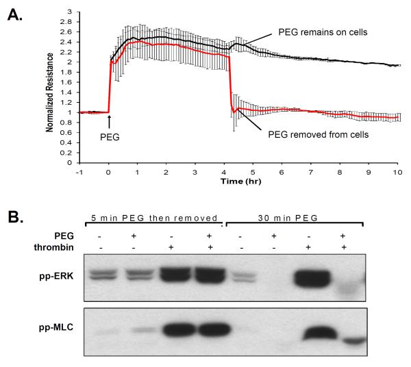
Figure 2. PEG is required to be physically present to induce barrier enhancement and to block thrombin-induced ERK and MLC phosphorylation.
Prior data on epithelial cells suggest that PEG actives cells even upon removal of PEG. The response of PEG removal is examined in human lung EC. A) The effect of PEG on TER is assessed by pretreating cells with PEG (8%, 4hrs) and then removal of PEG upon replacement with EGM2 as compared with cells in which PEG was left on the cells. B) To examine the effect of PEG on signal transduction, cells were either pretreated with vehicle or PEG (8%, 30 min), and then stimulated with thrombin (1U/ml, 5 min). In comparison, cells were also pretreated with PEG for 5 min followed by EGM2 replacement to remove PEG (25 min incubation), and subsequently stimulated with vehicle or thrombin. Cell lysate were processed via Western blots probed with either antibodies specific for phospho-ERK (Thr202/Tyr204) or phospho-MLC (Thr18/Ser19). The presence of PEG abolishes thrombin-induced ERK and MLC phosphorylation, but removal of PEG eliminates the inhibitory effects of PEG.

