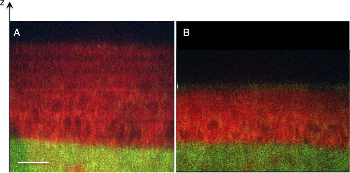
Figure 4.
Transverse/sagittal line scans of mouse cornea exposed to hypotonic saline. Albino mouse eyes (FVB) were exposed to either hypotonic saline (A) or isotonic saline (B) for 1 hour prior to multiphoton imaging. CARS signal shown in red and TPAF shown in green. The cells of the corneal EP appear swollen after treatment with hypotonic saline (A).

