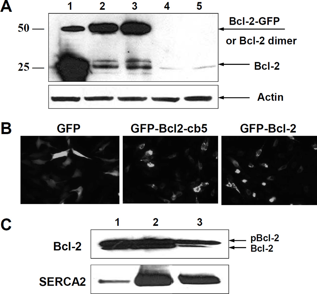
Figure 1. Expression of Bcl-2 in C2C12 myoblast cells.
A – C2C12 myoblast cells were harvested after 24 h of transfection with different Bcl-2 DNA chimeras and the level of Bcl-2 protein expression was analyzed by WB: 1- Bcl-2-cb5; 2 – GFP-Bcl-2; 3 – GFP-Bcl-2-cb5; 4 – GFP; 5 – empty vector. B – Fluorescent microscopy images of the C2C12 cells after overexpression of GFP (left), GFP-Bcl-2-cb5 (middle), and GFP-Bcl-2 (right). C – C2C12 myoblast cells were transfected with Bcl-2 DNA, and after 24 h, fractionated by differential centrifugation and analyzed by WB for Bcl-2 and SERCA2 in: (1) whole cell lysate, (2) mitochondria, and (3) microsomal fraction.

