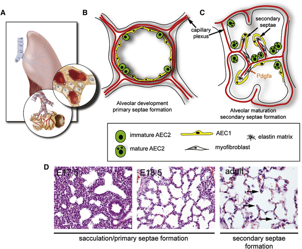
Figure 4. Schematic of Alveolarization.
(A) Alveolar development begins in late gestation as the endothelial plexus becomes tightly associated with the distal epithelial saccules. Bottom panel shows terminal bronchioles with saccules attached while top panel is a higher magnification after the saccules have been removed to reveal the lumen.
(B) The epithelium of the developing saccules/primary septae differentiates into several important cell types found in the mature alveolus. These include AEC1 cells, which are very flat and thin walled and when mature characteristically express aquaporin 5 and T1alpha (podoplanin), and the much larger, cuboidal AEC2 cells. These cell types are closely juxtaposed to each other and AEC1 cells form intimate interactions with the underlying vascular endothelium.
(C) Mature AEC2 cells secrete abundant surfactant proteins and lipids that are trafficked to the cell surface in organelles called lamellar bodies marked by the ACBC3 transporter protein. Mature AEC2 cells express high levels of the gene encoding surfactant protein C (Sftpc). Maturation of the alveolar compartment is accompanied by generation of secondary septae, which involves growth of alveolar crests. Crest formation requires elastin deposition and Pdgfa for myofibroblast development (Boström et al., 1996).
(D) Hand Estained histological sections demonstrating the change in distal lung morphology from the canalicular/saccular stages at approximately E17.5 through the adult where mature alveoli are found with secondary septae (arrows).

