Figure 9.
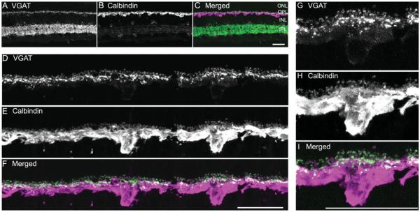
VGAT immunoreactivity is localized to horizontal cell bodies and processes. A vertical section through an adult guinea pig retina was double immunostained with antibodies to VGAT and calbindin. A: VGAT immunoreactivity is present in the cell bodies and processes in the OPL; VGAT immunoreactivity is distributed to amacrine cell and displaced amacrine cell somata and their processes in the IPL. B: Calbindin is expressed by horizontal cell somata and processes in the outer retina, and some amacrine and ganglion cell somata and their processes in the inner retina. C: Merged image shows the localization of VGAT and calbindin immunoreactivities in the retina. D–F: Enlarged images of the OPL show the co-localization of VGAT and calbindin immunoreactivities in horizontal cells. D: VGAT-immunoreactive processes and tips in the OPL. E: Calbindin-immunoreactive horizontal cell bodies and processes. F: Merged image shows the co-localization of VGAT and calbindin immunoreactivities in the OPL, indicating that VGAT immunoreactivity is localized to horizontal cell bodies and processes. G–I: Enlarged images show distribution of VGAT and calbindin immunoreactivities in a horizontal cell. G: Weak VGAT immunoreactivity is localized at the cell body, whereas strong VGAT immunoreactivity is distributed to processes and tips. H: Calbindin immunoreactivity is in horizontal cell body, processes, and tips. I: Merged image indicating that VGAT immunoreactivity is in horizontal cell body and processes, with more intense immunoreactivity in the tips. Confocal images were scanned at 1-μm intervals, and six optical sections were obtained and compressed for viewing. Scale bar = 20 μm in C (applies to A–C), F (applies to D–F), and I (applies to G–I).

