Figure 2.
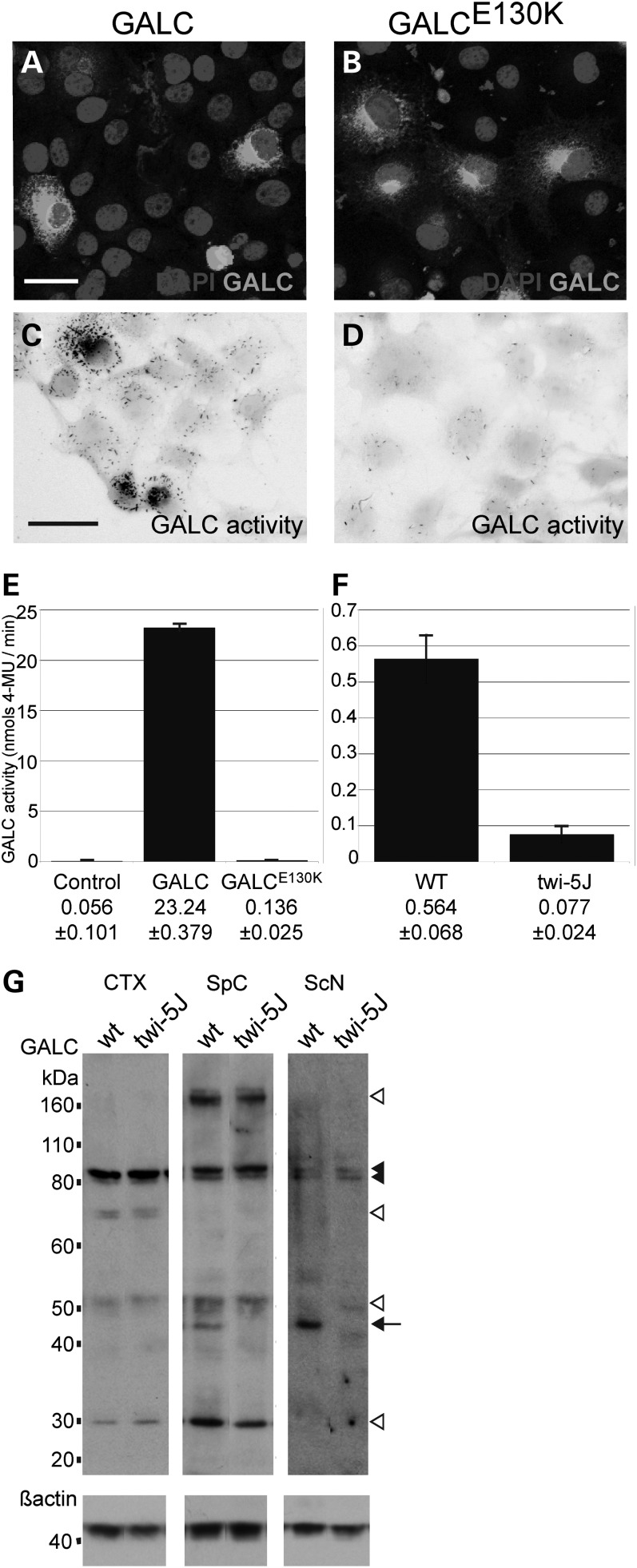
The E130K substitution in GALC abolishes GALC enzymatic activity without affecting precursor protein levels in vivo. (A–D) COS-7 cells were transfected with an expression construct for GALC (A and C) or GALCE130K (B and D). (A and B) GALC (white) was detected by immunofluorescence using a GALC-specific antibody. (C and D) GALC activity (black) was visualized using a GALC histochemical assay. Cell bodies are false-colored grey. Scale bar in A, C = 50 µm. (E) GALC activity (nmols/min) was measured from 15 µg of COS-7 cells extracts transfected with expression constructs for wild-type GALC or GALCE130K. Controls are mock-transfected cells. (E) GALC activity (nmols/min) measured from 15 µg of whole brain extracts from WT and twi-5J. Activity is presented as mean ± SD. (G) Western blot analysis using an affinity purified GALC antibody on extracts from wild-type (wt) and twi-5J collected from cortex (CTX), spinal cord (SpC) and sciatic nerve (ScN). Expression of ∼80, ∼50 and ∼30 kDa GALC bands are equivalent in cortical extracts between wild-type and twi-5J (P > 0.05). In contrast, a ∼45 kDa band is absent in extracts collected from mutant spinal cord and sciatic nerve compared with wild-type controls (P < 0.001) (arrow). The ∼80 kDa GALC precursor (black arrowheads) is present in all tissues. White arrowheads point to GALC species differentially regulated between tissues. Blots were re-probed using a beta-actin antibody to control for protein loading. Apparent molecular mass is indicated on the left. Extracts from three independent wild-type and twi-5J animals were analyzed with equivalent results; a representative immunoblot is shown.

