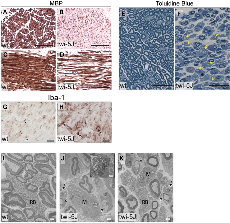
Figure 9.
The sciatic nerves of twi-5J are severely hypomyelinated with macrophage accumulation. (A–D) MBP immunohistochemistry on cross-sections (A and B) and transverse-sections (C and D) through the sciatic nerve of P23 wild-type (A and C) and twi-5J (B and D) indicate significant myelin-deficiency in the PNS. (E and F) Toluidine blue staining on thin sections through the sciatic nerve of P25 wild-type control (E) and twi-5J (F). Compared with wild-type, the twi-5J sciatic nerve is less compact, exhibits ∼22% loss of axons and contain numerous hypomyelinated axons (arrowheads), macrophages (M) and large lipid-filled vesicles (arrows). (G and H) Iba-1 immunohistochemistry on cross-sections reveal accumulation of globoid cells (arrows) in twi-5J. Iba-1 also detects non-reactive macrophages in control and twi-5J sciatic nerve (arrowhead). (I–K) Electron microscopy of sciatic nerve of twi-5J reveals numerous macrophages, hypomyelinated axons and lipid inclusions compared with wild-type. Inset in J shows globoid cell. Black Arrows point to lipid inclusions. Arrowheads indicate hypomyelinated axons. M, macrophage; RB, Remak bundle. Scale bars: A–D = 250 µm; E–H = 25 µm; I–K = 1 µm.

