Figure 6.
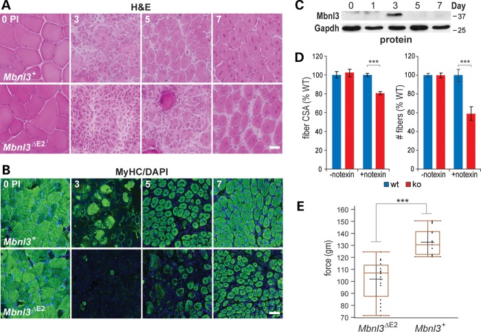
Impaired muscle regeneration in Mbnl3ΔE2 knockouts. (A) H&E (scale bar = 50 μM) and (B) MyHC (scale bar = 100 μM) immunofluorescence of transverse TA muscle sections from Mbnl3+ and Mbnl3ΔE2 8-month-old mice before (0) or 3, 5 and 7 days post-notexin injection to induce muscle regeneration. (C) Immunoblot confirming that the Mbnl3ZnF1-4 protein is detectable at day 3 PI during TA regeneration of older (8-month old) Mbnl3+ wild-type mice. (D) Bar graphs showing that the fiber cross-sectional area (left) and fiber number (right) in Mbnl3ΔE2 knockouts (red) are significantly reduced compared to wild-type (blue) following notexin treatment (+notexin) but are not affected in muscles in the absence of notexin (-notexin). (E) Grip strength assay indicating average force exerted by forelimb muscles in wild-type (Mbnl3+) versus Mbnl3ΔE2 mutant mice. Data in (C) and (D) are SEM (n = 3) and significant (***P < 0.001).

