
Figure 8.
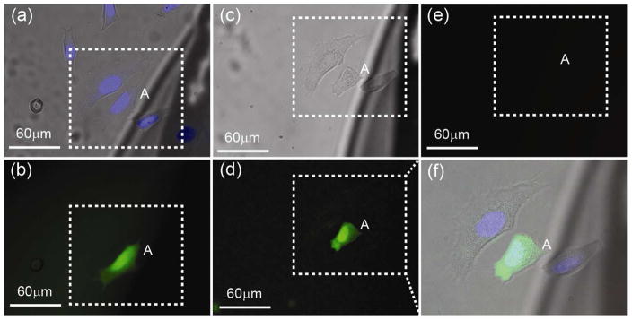
Live/dead assay using propidium iodide (PI). (a) Alexa Fluor was electroporated into target cell A. (b) Shows the corresponding fluorescence image. After electroporation, the coverslip was kept in an incubator for 4 hours and then stained with PI to image dead cells. (c)–(e) are bright field, green (Alexa Fluor), and red (PI) florescence, respectively, after PI staining. No red fluorescence was observed in (e), which indicates all cells in the field of view were alive. Note that (f) is the merged bright field and fluorescence zoom-in images in the region of interest (dotted box) after 4 hour incubation.