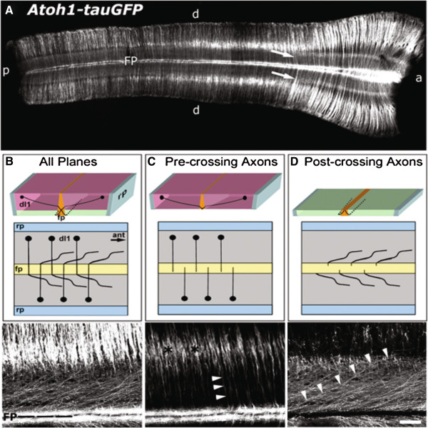
Figure 3.
Pre-crossing and post-crossing segments of GFP-labeled commissural axons visualized in distinct optical planes. (A) GFP immuno-labeling of an open-book preparation spanning the thoracic (left) to cervical (right) spinal cord derived from an E11.5 Atoh1-tauGFP mouse. Given the bilateral symmetry of the spinal cord, commissural axons from each side cross the ventral midline and obscure the visualization of axons arising from neurons located on the opposite side. (B) The top panel is a three-dimensional drawing of the open-book and the middle panel is a two-dimensional schematic representing the view from above the spinal cord. The bottom panel represents a confocal micrograph (Z-stack) of all planes from the labeled open-book displayed in A, including pre-, midline-, and post-crossing segments of dI1 commissural axons. The labeling on each side of the FP contains post-crossing axons that project rostrally, immediately adjacent to the ventral midline. (C) The three- (top) and two-dimensional (middle) schematics depict the optical planes of the open-book that contain mainly dI1 commissural cell bodies and the pre-crossing axonal segments. The micrograph (bottom) is from the same region imaged in B that includes only optical planes containing cell bodies (black asterisks) and pre-crossing axons (arrowheads). (D) The three- (top) and two-dimensional (middle) schematics depict the optical planes (marginal zone of spinal cord) of the open-book that mainly contain post-crossing dI1 commissural axon segments. The micrograph (bottom) is from the same region examined in B and C, but includes only planes representing the most marginal surface of the open-book containing post-crossing axon segments (arrowheads outline one such axon). Scale bar in D, 500 μm for A, and 100 μm for B-D.

