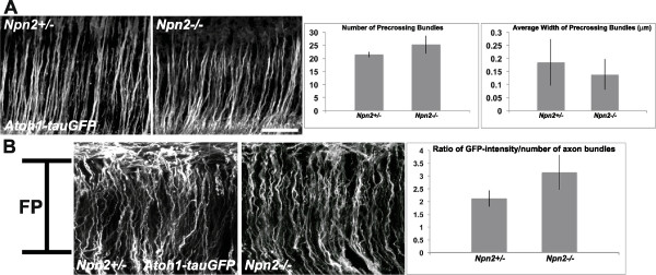
Figure 4.

The number of pre- and midline-crossing dI1 axons is unaltered in the absence of Neuropilin 2 (Npn2). Confocal micrographs from Z-stack optical planes that contain exclusively pre-crossing (A) or midline crossing (B) segments of labeled dI1 axons in open-book preparations derived from E11.5 Npn2−/+ or Npn2−/− mouse embryos harboring the Atoh1-tauGFP reporter. (A) The numbers of pre-crossing axon bundles were counted in images representing three separate pre-crossing axon-containing regions from the Npn2 null or heterozygous littermate embryos. There was no statistical difference between the numbers of axon bundles in homozygous and heterozygous embryos (Student’s T-test). The width (in μm) of individual bundles was measured for the same sets of images used to calculate the numbers of axon bundles and there was no statistically significant difference (Student’s T-test) between the values obtained for Npn2 null mouse embryos and their littermate controls. (B) The average GFP-intensity of each section was normalized to the number of axon bundles crossing the ventral midline at the FP to generate a ratio of GFP-intensity/axon bundles from three separate FP-containing regions of a given Npn2 null embryo and compared with the measurements obtained from heterozygous littermate control embryos. A Student’s T-test showed no statistically significant difference between the numbers of axons in the homozygous or heterozygous preparations. The data are derived from 4 to 5 embryos per genotype, per measurement. Scale bar = 100 μm in A, and the width of the FP in B (see bar in left-most panel).
