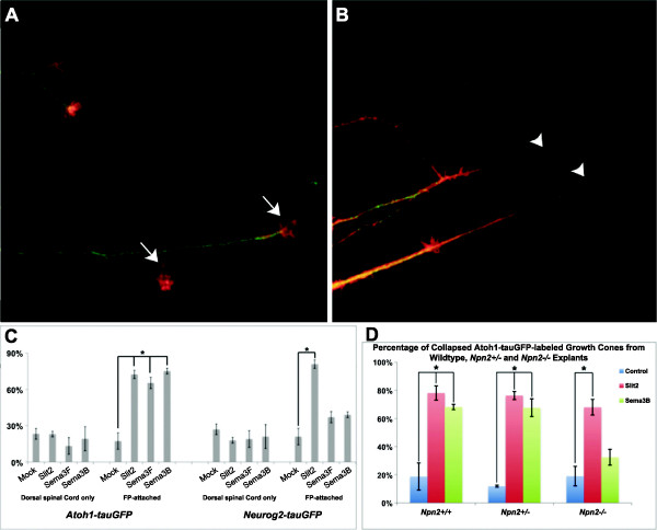
Figure 7.
Sema3B or Sema3F collapses growth cones associated with post-crossing dI1, but not dI4, axons. Floor plate (FP)-attached (post-crossing) and dorsal spinal cord only (pre-crossing) explants containing dorsal commissural neurons were generated from open-book preparations of E11-E11.5 Atoh1-tauGFP(A, B) or Neurog2-tauGFP (data not shown) spinal cords. (A) Growth cones (GCs) from a post-crossing explant derived from an Atoh1-tauGFP open-book and labeled with Phalloidin (red) and GFP (green) remained well spread after 1 h mock-conditioned medium treatment. (B) Many of these GCs collapsed (absence of lamellipodia/filopodia) when treated for 1 h in media conditioned with Slit2, Sema3F or Sema3B. (C) The percentage of collapsed GCs associated with Atoh1-tauGFP (dI1) or Neurog2-tauGFP (dI4) pre-crossing axons treated with Slits-, Sema3F- or Sema3B-conditioned media was not significantly different as compared to mock. A significant increase in the percentage of collapsed GCs was observed with post-crossing dI1 axons treated Slit2, Sema3F or Sema3B, as compared to mock-media (chi-squared test, P value <0.05). No difference in the percentage of collapsed GCs was observed with post-crossing dI4 axons in the presence of Sema3B or Sema3F, as compared to mock-media. A significant increase in the collapse of dI4 axon-associated GCs was observed when treated with Slit2 as compared to mock-media (chi-squared test, *P value <0.05). (D) Growth cones from Npn2+/+ and Npn2+/− post-crossing axons displayed significant increases (chi-squared test, *P value <0.05) in the percentage of collapse when treated with Slit2 or Sema3B, as compared to mock-media. Sema3B failed to induce a significant increase over mock in the percentage of collapsed GCs from Npn2−/− embryos. The data are derived from 4 to 5 embryos/genotype. The percentage of collapse was calculated as the number of collapsed GCs divided by the total number of GCs. ±50 GCs were analyzed for each condition and in each experiment.

