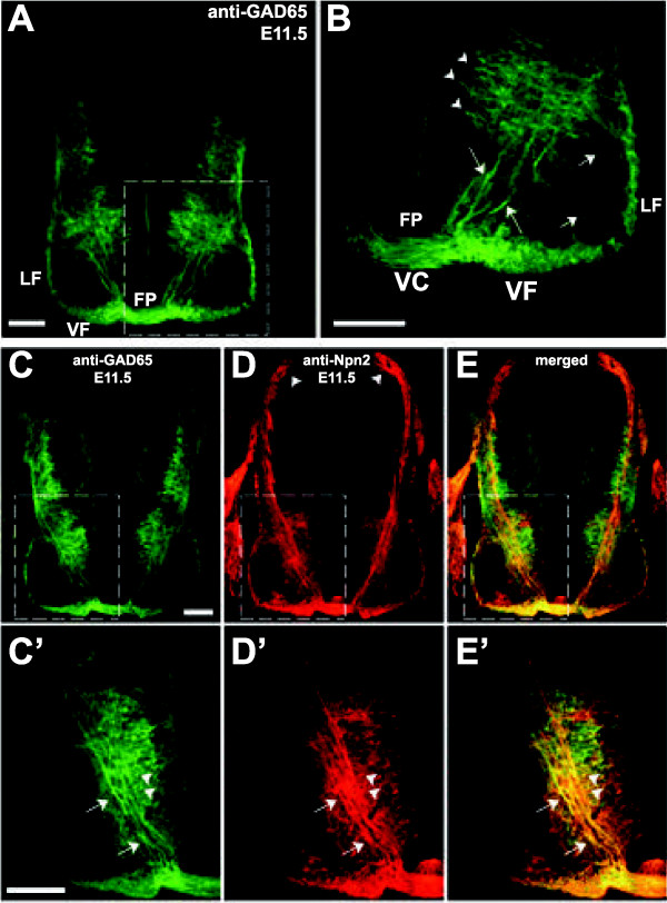
Figure 8.
Ventral spinal commissural neurons are GABAergic and express Neuropilin 2 (Npn2). (A-B), A wild type E11.5 mouse spinal cord transverse section is labeled with anti-GAD65 to visualize the large population of GABAergic commissural neurons (CNs) with cell bodies (arrowheads) located in the ventromedial region of the intermediate zone (dashed box area is shown at high magnification in B). Axons (arrows in B) emanating from ventromedial GABAergic CNs project ventrally toward the floor plate (FP) and cross the midline within the ventral commissure (VC), and then turn rostrally to join the ventral funiculus (VF). Some GAD65-positive neurons also extend their axons ipsilaterally (shorter arrows in B) to join the lateral funiculus (LF). (C-E’), Some GAD65-positive CNs from the ventromedial population also express Npn2 on the ipsi- and contralateral segments of their axons as illustrated by the double immunofluorescence (E, merge) labeling of a single spinal cord transverse section with anti-GAD65 (C, green) and anti-Npn2 (D, red). Higher magnification of the dashed boxed areas in the single (C-D) or double/merge (E) labeled section are shown in C’-E’, and reveal that a subset of the ventromedial GABAergic CNs expresses both GAD65 and Npn2 on their axons (arrows) and cell bodies (arrowheads). Scale bars in A and C, 50 μm for A, C-E; in B and C’, 100 μm for B, C’-E’.

