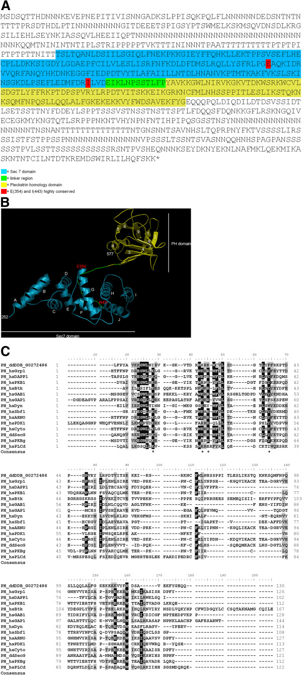
Figure 1.
Domain structure of the Sec7 protein. (A) The amino acid sequence of Sec7 of D. discoideum is shown (DDB0233617). The Sec7 domain is highlighted in blue, the linker region in green and the Pleckstrin homology (PH) domain in yellow. The highly conserved amino acids E(354) and I(443) are marked with red. (B) The DDB0233617 Sec7-PH-domain modeled to 2r0dA (Crystal Structure of Autoinhibited Form of Grp1 Arf GTPase Exchange Factor). The Sec7 domain is depicted in blue, the linker region in green and the PH domain in yellow. The highly conserved amino acids E(354) and I(443) are shown in red. Modeling was with SwissModel, visualization with OpenAstexViewer. (C) CLUSTALX alignments of PH domains from Homo sapiens and D. discoideum according to Lietzke et al. and Ferguson et al. [11,12]. The asterisks in the Consensus line indicate the signature motif as suggested by Lietzke et al. and Ferguson et al. [11,12]. ddDDB_G0272486 corresponds to Sec7.

