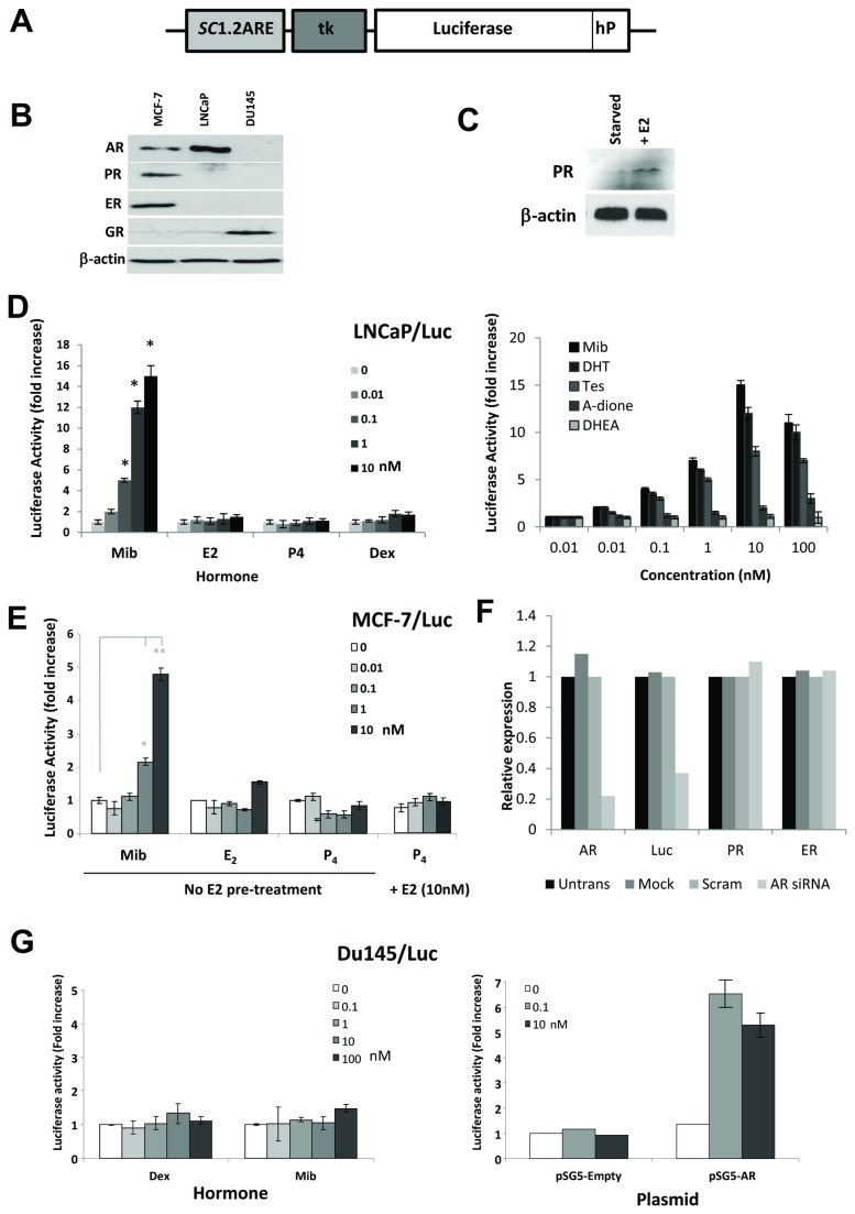
Figure 1. Analysis of the androgen receptor reporter construct in stably transfected cells.
(A) Schematic representation of the ARE-tk-Luc androgen receptor reporter construct (hP = hPEST degradation signal). (B) Western blot analysis of steroid receptor expression in MCF-7, LNCaP and Du145 cells growing in full media. (C) Western blot analysis of PR expression in hormone starved MCF-7 cells -/+ E2 treatment for 24hr. (D), Luciferase activity from hormone treated LNCaP/Luc cells – treated for 24hours with 0-10nM mibolerone (Mib), estrogen (E2), progesterone (P4) and dexamethasone (Dex) (left hand side) and with 0-100nM of the androgens - (Mib), dihydrotestosterone (DHT), testosterone (Tes), androstenedione (A-dione) and dehydroepiandrosterone (DHEA) for 24 hours (right hand side). (E) Luciferase activity from hormone treated MCF-7/Luc cells – treated for 24hours with 0-10nM Mib, E2 or P4 or with additional 24hr pre-treatment with E2 (10nM) to induce PR expression. (F) Q-PCR quantification of relative expression of the steroid receptors AR, ER, and PR and luciferase transcripts in MCF-7/Luc cells, grown in full medium, transfected with siRNA against androgen receptor. (F) Luciferase activity from Du145/Luc cells treated with 0-100nM dex or mib (left hand side) or pre-transfected with an additional AR or empty vector expression constructs (right hand side). **P<0.01, *P<0.05 (t-test analysis).

