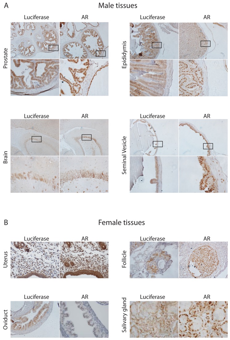
Figure 6. Analysis of AR and luciferase expression within a sample of mouse tissues.
(A), Immunohistochemical co-localisation staining for AR (nuclear and cytoplasmic_ and luciferase (cytoplasmic) on consecutive formalin-fixed tissue sections taken from the ARE-Luc male mice. Upper panel represents image at 10x magnification, with box inset and lower panel showing 40x magnification. (B), Immunohistochemical staining for AR in a variety of female mouse tissues.

