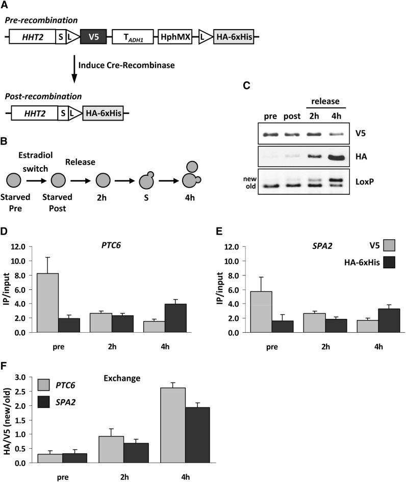
Figure 7.
Immunoblot and ChIP analysis of histone H3 dynamics. (A) Histone H3 was tagged with a V5→HA-6xHis RITE cassette in strain NKI8050. (B) Outline of experimental setup. (C) Immunoblot analysis of cells arrested by starvation (pre), switched for 16 hr (post), and subsequently released into fresh media for 2 hr (no cell division) or 4 hr (one cell division). An antibody against spacer-LoxP detects both old and new H3. (D-E) H3-V5 and H3-HA-6xHis ChIP analysis of samples described in (B) to measure occupancy (IP/input) of old and new H3 at PTC6 (around transcription start site) and SPA2 (coding sequence) (primer sequences are listed in Table 3). (F) Exchange of histone H3 determined as the ChIP ratio of new H3 over old H3 (HA/V5). Average of three biological replicates ± SEM.

