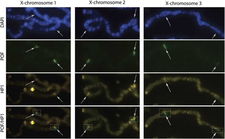
Figure 4.
POF and HP1a binding overlap on the PoX loci. POF (green) and HP1a (yellow) on the X-chromosome in three representative female nuclei. The proximal parts of the X chromosomes are shown, and the PoX1 and PoX2 loci are indicated with arrows. The boxes in the POF/HP1 row show the combined image. Note that levels of HP1a binding at PoX loci are above the background staining level.

