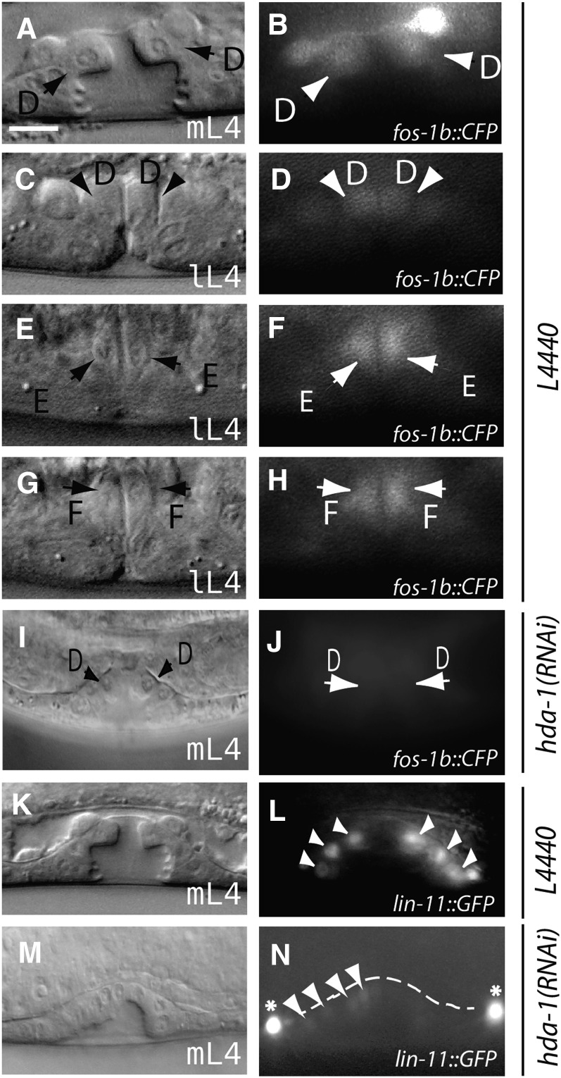
Figure 3.
lin-11 and fos-1 expression is altered in hda-1 mutants. DIC and corresponding fluorescent images of animals expressing a translational fos-1::cfp reporter. (A and B) Control L4440 RNAi-treated mid-L4 animal showing fos-1 expression in presumptive vulD cells. (C−H) mid/late-L4 stage animals showing fos-1 expression in presumptive vulD, vulE and vulF cells. (I, J) hda-1 knockdown causes reduction in fos-1::cfp expression. Diffuse CFP fluorescence is observed in the region overlapping with presumptive vulD cells. lin-11 expression is detected in vulval cells in control RNAi-treated animals (K, L) but is absent in hda-1-RNAi treated animals (M, N). Some of the GFP fluorescing cells are marked by arrowheads and arrows (D, E and F refer to vulD, vulE and vulF, respectively). mL4: mid-L4, lL4: late-L4. Asterisk in panel N points to VC neuronal cells. Scale bar is 10 μm.

