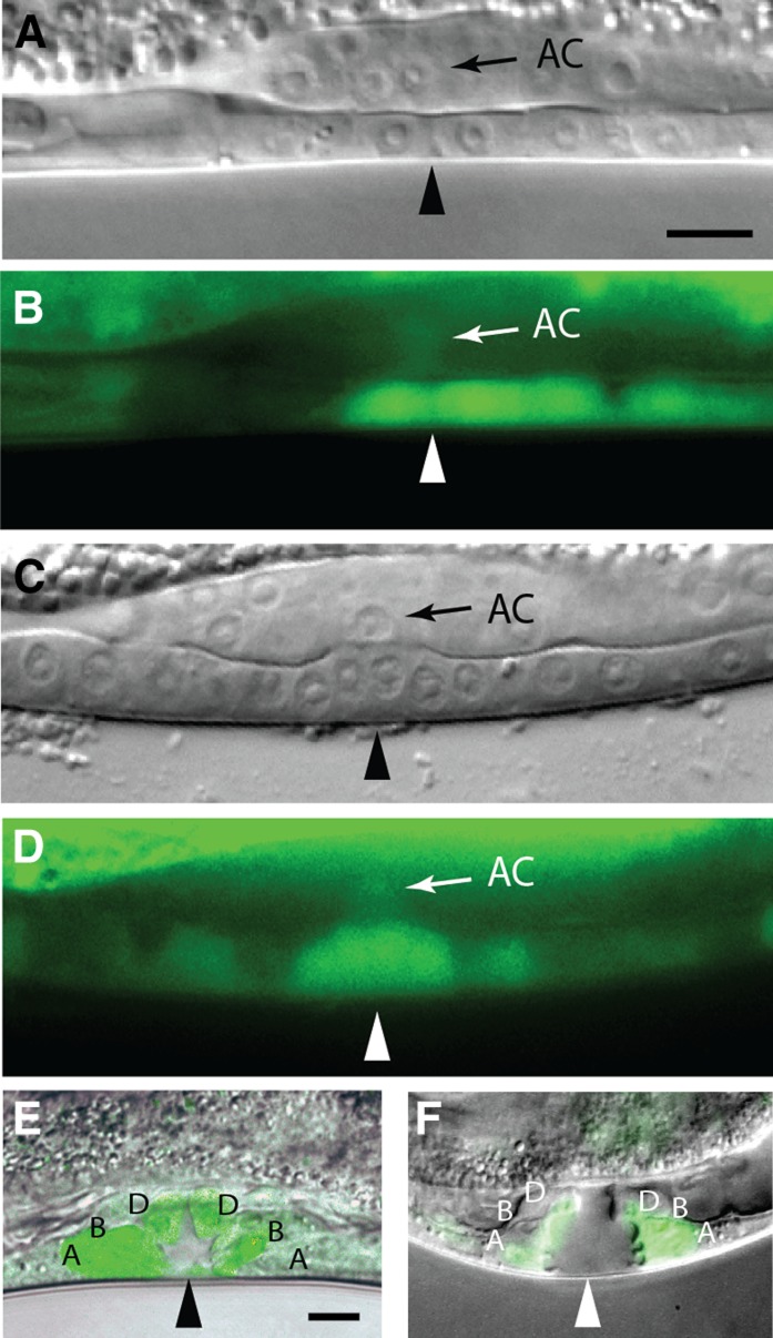
Figure 4.
hda-1 expression in the vulva and AC. (A−E) sEx13706 and (F) bhEx68. (A, B) Pn.px cells. (C, D) Pn.pxx cells. (E, F) Pn.pxxx cells. Triangles mark the center of vulval invagination. The presumptive vulval cell types A (vulA), B (vulB1 or vulB2), and D (vulD) are shown. The AC is shown with arrows. In (B), P5.p is in the process of dividing and has reduced level of GFP fluorescence. Scale bar is 10 μm.

