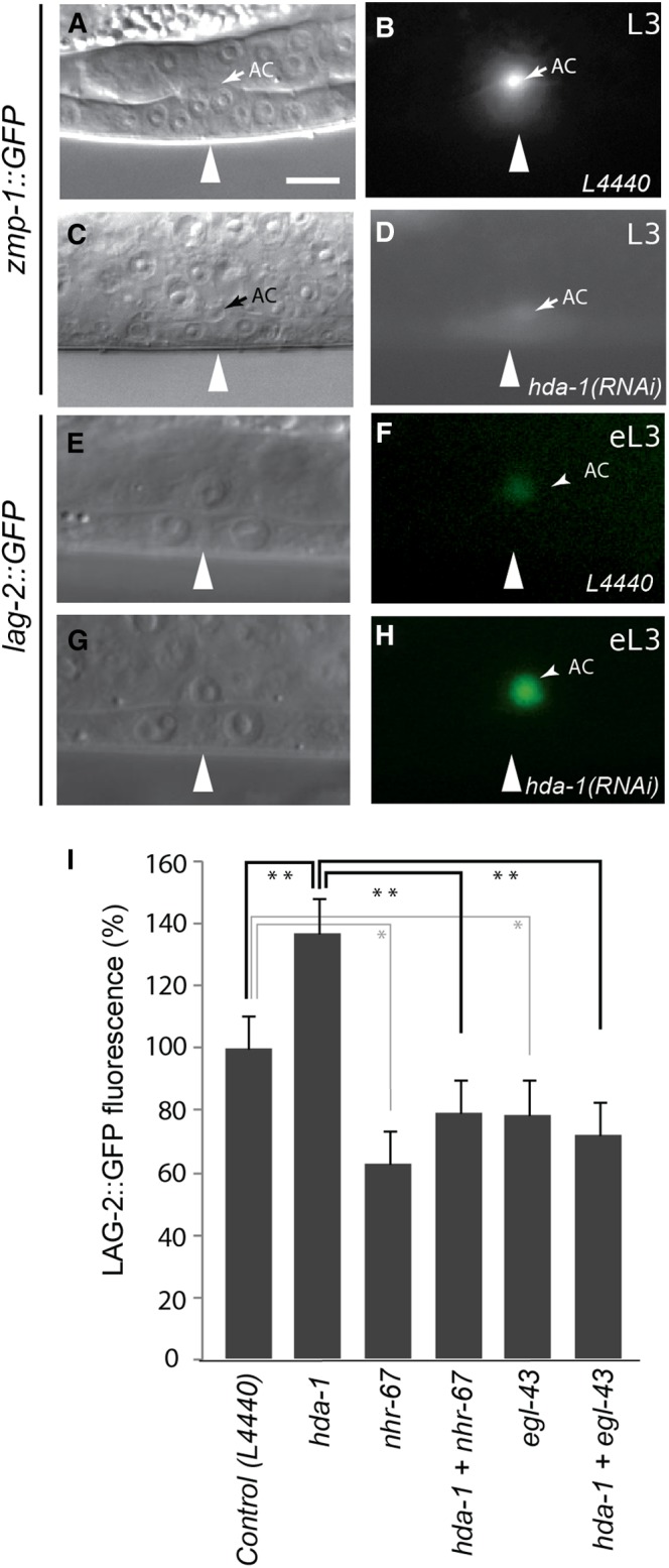
Figure 7.

Effect of hda-1 RNAi knockdown on the AC. (A, B) zmp-1::gfp expression in the AC of a wild-type animal. (C, D) zmp-1 expression is strongly diminished in hda-1(RNAi) animal. (E, F) Wild-type lag-2::gfp (arEx1352) expression in the AC. (G, H) GFP fluorescence in AC is brighter in hda-1(RNAi) animal. Arrowheads mark the center of vulval invagination. Scale bar is 10 μm. (I) Quantification of lag-2::gfp fluorescence intensity in the AC. The hda-1(RNAi) animals show a significant increase in GFP fluorescence compared with controls. In contrast, nhr-67(RNAi) and egl-43(RNAi) animals show reduced GFP fluorescence in the AC. The increase in lag-2::gfp fluorescence in hda-1(RNAi) animals was suppressed by nhr-67(RNAi) and egl-43(RNAi). 20 or more animals were examined in each case. eL3, early-L3. The P values for pairs are indicated by stars (**P < 0.01, *P < 0.05).
