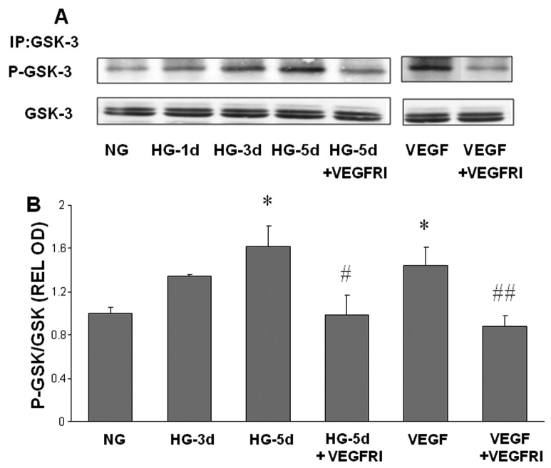
Figure 4. High glucose-induced phosphorylation of GSK3β.
Endothelial cells were grown in serum-free medium containing normal glucose (NG, 5.5 mM) or high glucose (HG, 25 mM) with or without VEGFR inhibitor (VEGFRI) for 1 to 5 days. Equal amounts of SDS extracted protein samples were immunoprecipitated (IP) by anti-GSK3β antibody and subjected to SDS-PAGE and Western blotting (A). Densitometric analysis of phospho-GSK3β bands, normalized for the corresponding GSK3β bands, showed that phosho-GSK3β levels were significantly increased by HG or VEGF treatment and that this effect was reduced in VEGFI treated samples compared to HG or VEGF treated endothelial cells (B). * = P<0.05 vs NG, # = P<0.05 vs HG5, # # = P<0.05 vs VEGF.

