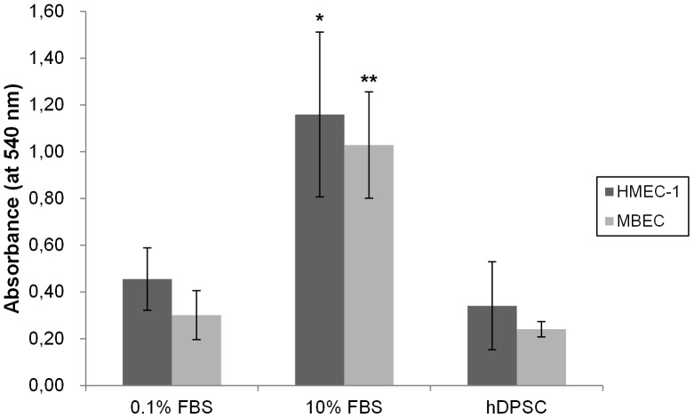
Figure 3. Cell proliferation (MTT) assay: MBEC or HMEC-1 endothelial cells were seeded in 96-well plates and 24 hours later the CM of hDPSC was added.
αMEM containing 10% FBS was included in the study as a positive control. 72 h later, the media were removed and cell proliferation was assessed. Conditioned medium of hDPSC was not able to increase the cell growth of neither MBEC nor HMEC-1 compared to control medium (with 0.1% FBS). Incubation of αMEM with 10% FBS resulted in a 3-fold increase in cell growth in both MBEC and HMEC-1. This assay was repeated 3 times with a total number of at least six different donors; *p<0.05; **p<0.01.

