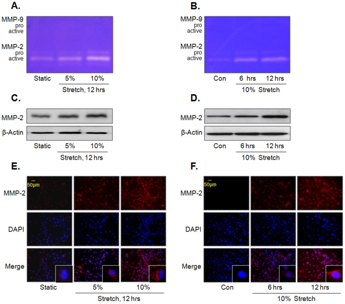
Figure 1. Effects of MS on MMP-2 activity and expression in VSMC.
Cells were stimulated by MS at indicated forces (A) or time (B), and then gelatinolytic activity (MMP-2 and -9) was determined using gelatin zymography. The force- and time-dependent increase in MMP-2 expression in VSMC exposed to MS was determined by Western blot (C and D) and immunocytochemical studies (E and F). Representative images are from 4–6 independent experiments.

