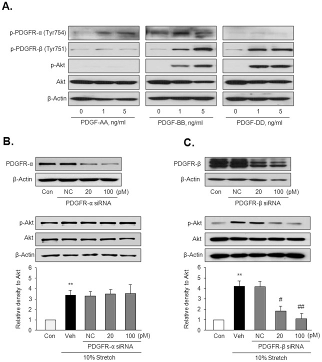
Figure 6. The individual roles of PDGFR isoforms in Akt phosphorylation in VSMC.
(A) Representative immunoblots for the phosphorylated (p-Akt) and total Akt in cells stimulated with a PDGFR-α ligand (PDGF-AA) and PDGFR-β ligands (PDGF-BB and –DD) for 10 min (n = 5). (B) Cells were transfected with the indicated doses of PDGFR-α siRNA or PDGFR-β siRNA, and then stimulated by 10% MS for 4 hrs. Quantitative data for the corresponding blots are presented as means ± SEM (n = 5). **p<0.01 vs. control (Con). #p<0.05, ##p<0.01 vs. vehicle (Veh).

