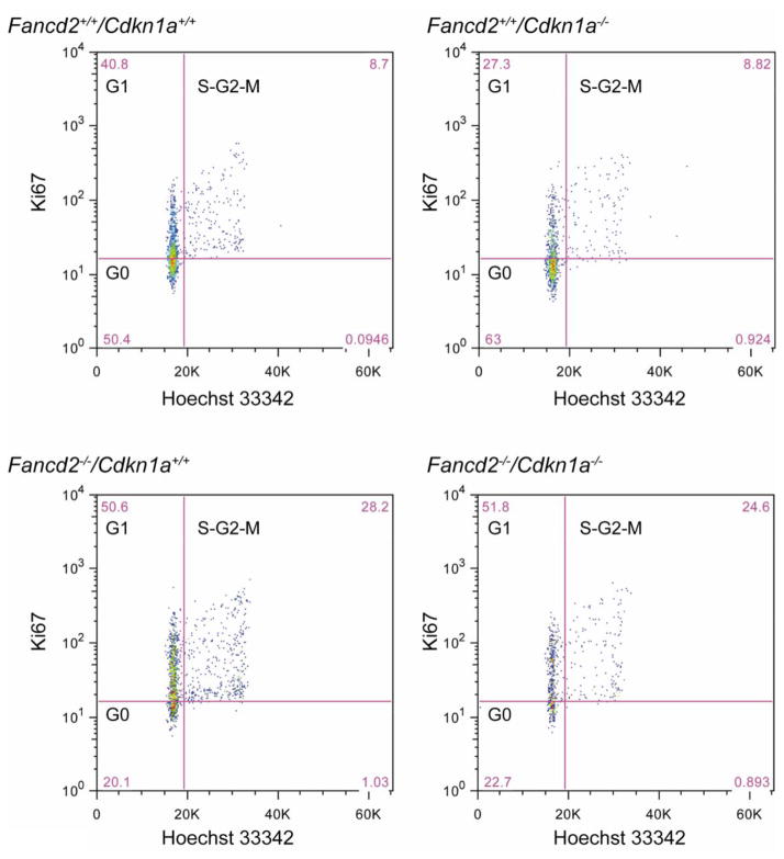
Figure 2. Representative flow cytometry profile of cell cycle analysis.
p21 deletion did not change the abnormal cell cycle status of Fancd2−/− mice. Hoechst 33342 and FITC-conjugated anti-mouse Ki67 (BD Sciences) were used in combination to distinguish cells in G0, G1, and S-G2-M phases of the cell cycle.

