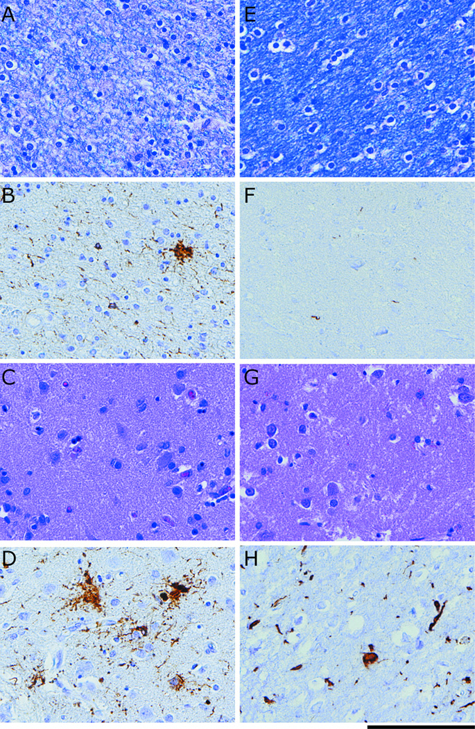
Figure 3.
Neuropathological evidence for a dissociation of grey matter (GM) and white matter (WM) burden in representative cases of FTLD-TAU and FTLD-TDP at 40x (Scale bar represents 100μm): (A) LFB stain for WM degeneration for FTLD-TAU; (B) Tau inclusions in WM for FTLD-TAU; (C) H-E stain for neuronal loss in GM for FTLD-TAU; (D) Tau inclusions in GM for FTLD-TAU; (E) LFB stain for WM degeneration for FTLD-TDP; (F) TDP-43 inclusions in WM for FTLD-TDP; (G) H-E stain for neuronal loss in GM in for FTLD-TDP; (D) TDP-43 inclusions in GM for FTLD-TDP.

