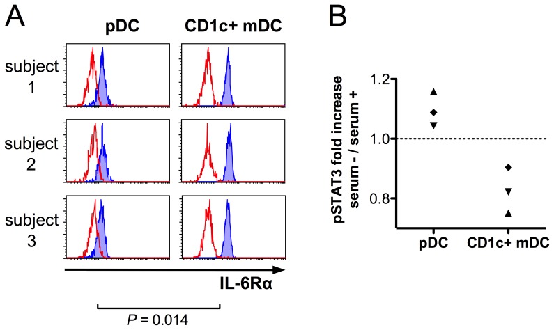
Figure 6. pDCs express cell-surface IL-6Rα and are not dependent on the presence of serum components for IL-6 signal transduction.
Flowcytometric analysis of IL-6Rα expression on DC subsets was performed in whole blood samples from healthy volunteers. IL-6Rα is expressed on pDCs, although in lower levels than on CD1c+ mDCs (A). PBMCs were isolated from fresh peripheral blood samples from three healthy volunteers in independent experiments. Cells were incubated at 37°C in RPMI 1640 culture medium (supplemented with 2 mM L-glutamine) either with 1/3 vol. of autologous serum or without serum for one hour prior to stimulation with 50 ng/ml of recombinant IL-6 for 10 minutes (in the presence or absence of serum), followed by flowcytometric DC identification and pSTAT3 measurement using similar gating strategy as indicated in Fig. 1. Despite lower IL-6Rα expression on pDCs, IL-6-induced pSTAT3 response in pDCs is not impaired in the absence of autologous serum, whereas mDCs (serving as intrinsic controls) exhibit somewhat reduced responses when stimulated in serum-free conditions. Samples from each donor are indicated with different symbols (B).

