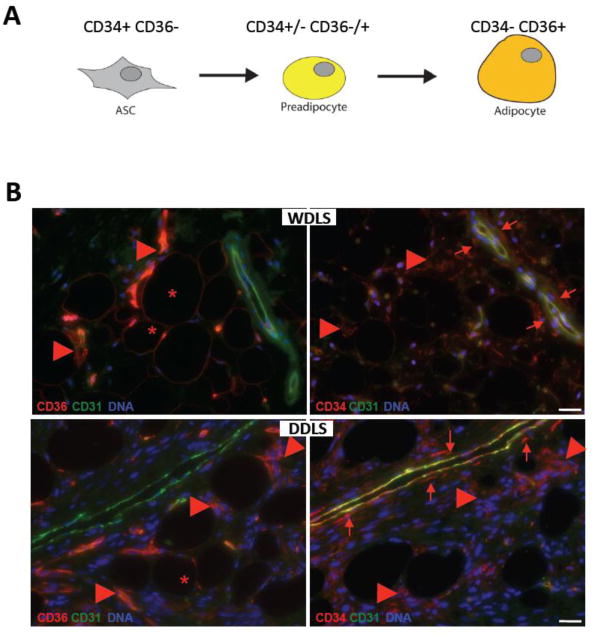
Figure 1.
WDLS and DDLS cells at distinct differentiation stages. (A) A schematic depicting expression of CD34 and CD36, cell surface markers used for liposarcoma cell classification, during differentiation of mesenchymal adipocyte progenitors. (B) Immunolocalization of distinct liposarcoma populations in serial paraffin sections of representative WDLS and DDLS samples subjected to immunofluorescence with antibodies against CD36 or CD34 (red). CD36+CD34- cells with adipocyte morphology (*), adjacent stromal CD36+CD34+ cells (arrowheads) and mainly perivascular CD36-CD34+ cells (arrows) are indicated relative to non-malignant vasculature expressing CD31 (green). Nuclei are blue. Scale bar: 50 μm.

