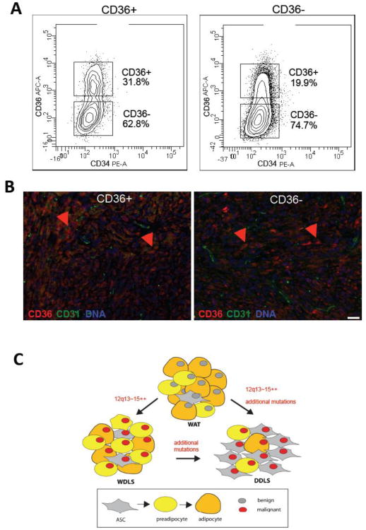
Figure 4.
Plasticity of liposarcoma populations in mouse xenografts. (A) Separation of cells from secondary xenografts of CD36+ (left) and CD36- (right) cells (Figure 3D-E) into CD36+ and CD36- populations. (B) Paraffin sections of xenografts grown from CD36+ cells (left) and CD36- cells (right) subjected to immunofluorescence with antibodies against CD36 (red) indicating comparable distribution of CD36-expressing cells relative to vasculature expressing CD31 (green). Nuclei are blue. Scale bar: 50 μm. (C) A model of liposarcomagenesis. Liposarcoma arises in benign WAT as a result of 12q13-15 amplification and MDM2 overexpression (red nucleus), which occurs at one of the stages of adipogenesis (box). DDLS arise either from WDLS though gradual dedifferentiation of progressive percentage of tumor cells to the ASC-like phenotype or independently of WDLS by separate mutations in WAT leading to a comparatively more aggressive domination of malignant ASC-like cells.

