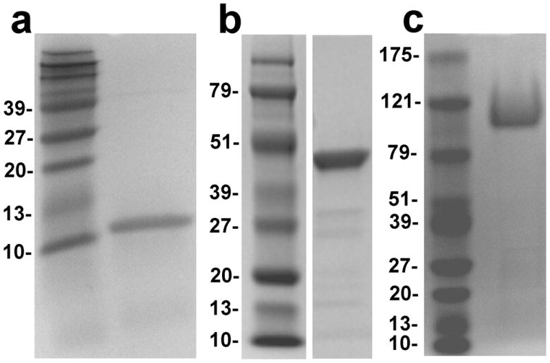
Figure 1.

SDS-polyacrylamide gel electrophoresis of the recombinant proteins. The GelCode Blue stained gels are shown. (a) Human colipase on an 18% gel; (b) Human PLRP2 on a 10% gel; (c) Human CEL on a 7.5% gel. Molecular weight markers are included in each panel and the size in kDal is given on the right edge of each panel.
