Figure 2.
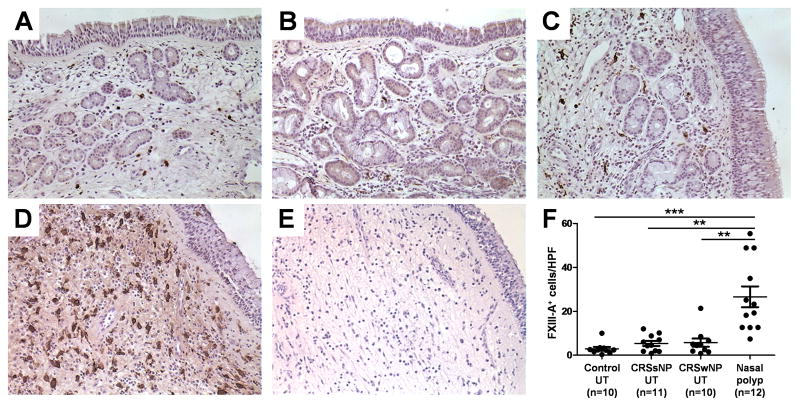
Immunohistochemistry of FXIII-A was performed with anti-human FXIII-A antibody. Representative immunostaining for FXIII-A in UT from control subject (A), a patient with CRSsNP (B), a patient with CRSwNP (C), and NP tissue (D). Negative control antibody staining in NP tissue from patient with CRSwNP (E) is shown. The number of FXIII-A+ cells in UT from control subjects (n = 10), patients with CRSsNP (n = 11), and patients with CRSwNP (n = 10) and NP (n = 12) was counted using a semiquantitative method (F). Magnification: ×400. ** p < .01, *** p < .001.

